
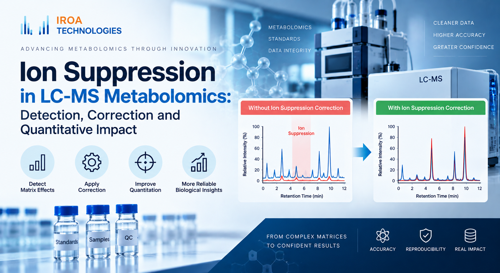
Ion suppression is a central analytical challenge in LC-MS metabolomics because measured signal intensity can decrease even when the true concentration of a metabolite has not changed. In complex biological matrices, co-eluting compounds may compete during ionization, changing response in a compound- and sample-dependent way. The result is a mismatch between abundance and observed peak area.
For quantitative work, this matters because uncorrected suppression can reduce sensitivity, increase variability, and create misleading differences between samples. IROA addresses this problem within the TruQuant Workflow Kit by using isotope-labelled internal standards and software-based suppression correction.
What Causes Ion Suppression?
In electrospray ionization, analytes must move from the liquid phase into gas-phase ions before they can be detected. Salts, phospholipids, highly abundant metabolites, extraction residues, and other matrix components can interfere with this process. The effect is commonly described as a matrix effect because the surrounding sample composition changes how efficiently a target compound forms detectable ions.
Suppression is often retention-time dependent. A metabolite may ionize efficiently in a clean standard solution but show weaker response in plasma, tissue, plant extract, or microbial samples because other compounds elute at the same time.
Why Raw Peak Area Can Be Misleading
A raw peak area represents instrument response, not concentration in isolation. If one sample experiences stronger suppression than another, comparing raw areas can overstate or understate biological differences. This is especially problematic in large batches where matrix composition and instrument conditions vary over time.
Quantitative metabolomics therefore benefits from an internal reference that experiences the same chromatographic and ionization environment as the analyte.
How Isotope-Labelled Internal Standards Help
Stable isotope-labelled internal standards are chemically similar to their natural-abundance counterparts but are distinguishable by mass. When a labelled signal and an unlabeled analyte co-elute, both experience the same local matrix environment. Their relationship can therefore reveal ionization losses that would be invisible from the analyte peak alone.
IROA’s internal standards use U-13C labelling to create identifiable isotope patterns across a broad metabolite mixture. The IROA Internal Standard Kit can be spiked into experimental samples so the reference signal is present during the same analytical run.
Suppression Correction In The Iroa Workflow
IROA uses the relationship between natural-abundance and isotope-labelled signals to estimate suppression effects and generate corrected quantitative values. This process is implemented in ClusterFinder, which supports isotope-pattern recognition, metabolite identification, ion suppression correction, and normalization.
Instead of treating suppression as an unavoidable source of noise, the workflow uses the internal standard to measure and correct part of the analytical loss. This is particularly useful when the goal is to compare metabolites across samples, batches, or chromatographic methods.
Suppression Correction And Normalization Are Different Steps
Suppression correction addresses local signal loss associated with ionization. Normalization addresses broader sample-to-sample or run-to-run differences. Both are needed for robust quantitative analysis because correcting one source of variability does not automatically correct the other.
After suppression correction, IROA applies Dual MSTUS normalization to improve comparability across samples. The combination is important because it separates compound-specific ionization effects from global analytical variation.
How To Recognize A Workflow That Needs Stronger Suppression Control
Warning signs include inconsistent response for the same sample type, reduced sensitivity in complex matrices, unexpected differences between replicate injections, retention-time regions with broad signal loss, and quantitative disagreement across batches or instruments.
Rather than relying only on post hoc statistics, a better strategy is to incorporate internal standards and QC materials during experimental design. IROA’s ion suppression correction resources and application notes provide additional technical context.
Conclusion
Ion suppression is not simply a signal-quality issue; it directly affects quantitative confidence. A workflow that measures suppression using isotope-labelled internal standards can distinguish analytical loss from biological change more effectively than raw-intensity analysis alone. Within TruQuant, suppression correction is integrated with identification, QC, and normalization to support more reproducible LC-MS metabolomics.